COPS3 Recombinant Monoclonal Antibody
-
中文名稱:COPS3重組抗體
-
貨號:CSB-RA556985A0HU
-
規格:¥1320
-
圖片:
-
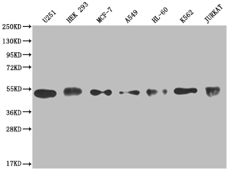 Western Blot
Positive WB detected in: U251 whole cell lysate, HEK293 whole cell lysate, MCF-7 whole cell lysate, A549 whole cell lysate, HL-60 whole cell lysate, K562 whole cell lysate, Jurkat whole cell lysate
All lanes: COPS3 antibody at 1:2000
Secondary
Goat polyclonal to rabbit IgG at 1/50000 dilution
Predicted band size: 48, 46 kDa
Observed band size: 40-55 kDa -
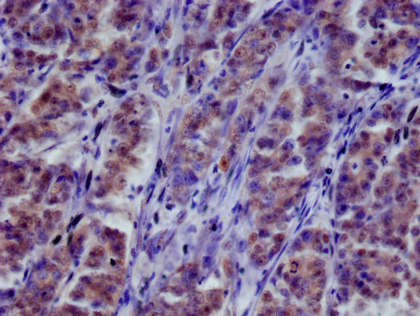
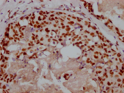
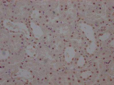
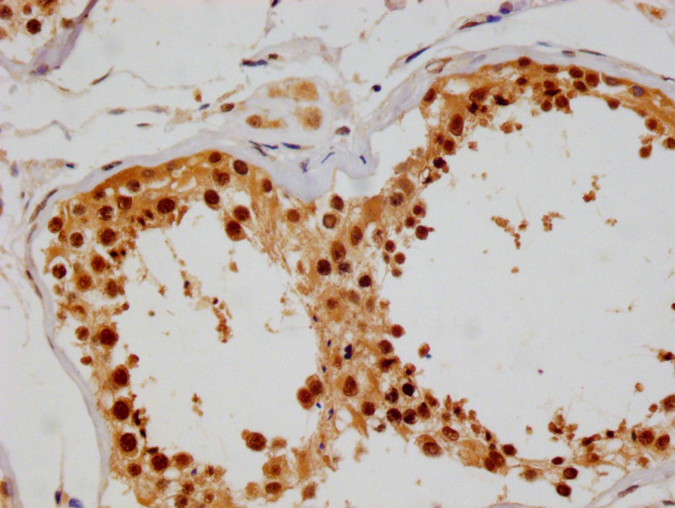 IHC image of CSB-RA556985A0HU diluted at 1:100 and staining in paraffin-embedded human testis tissue performed on a Leica BondTM system. After dewaxing and hydration, antigen retrieval was mediated by high pressure in a citrate buffer (pH 6.0). Section was blocked with 10% normal goat serum 30min at RT. Then primary antibody (1% BSA) was incubated at 4°C overnight. The primary is detected by a Goat anti-rabbit polymer IgG labeled by HRP and visualized using 0.05% DAB.
 -
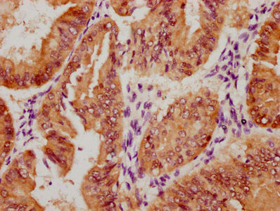
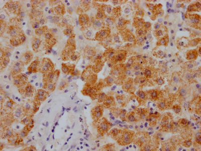
 IHC image of CSB-RA556985A0HU diluted at 1:100 and staining in paraffin-embedded human colon cancer performed on a Leica BondTM system. After dewaxing and hydration, antigen retrieval was mediated by high pressure in a citrate buffer (pH 6.0). Section was blocked with 10% normal goat serum 30min at RT. Then primary antibody (1% BSA) was incubated at 4°C overnight. The primary is detected by a Goat anti-rabbit polymer IgG labeled by HRP and visualized using 0.05% DAB.
 -
 Immunofluorescence staining of A549 cell with CSB-RA556985A0HU at 1:50, counter-stained with DAPI. The cells were fixed in 4% formaldehyde and blocked in 10% normal Goat Serum. The cells were then incubated with the antibody overnight at 4°C. The secondary antibody was Alexa Fluor 535-congugated AffiniPure Goat Anti-Rabbit IgG(H+L).
 -
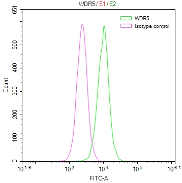
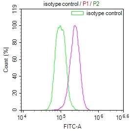
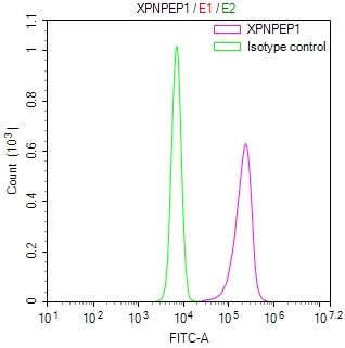
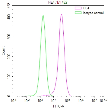
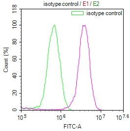 Overlay Peak curve showing MCF7 cells stained with CSB-RA556985A0HU (red line) at 1:100. The cells were fixed in 4% formaldehyde and permeated by 0.2% TritonX-100. Then 10% normal goat serum to block non-specific protein-protein interactions followed by the antibody (1ug/1*106cells) for 45min at 4℃. The secondary antibody used was FITC-conjugated Goat Anti-rabbit IgG(H+L) at 1:200 dilution for 35min at 4℃.Control antibody (green line) was rabbit IgG (1ug/1*106cells) used under the same conditions. Acquisition of >10,000 events was performed.
-
-
其他:
產品詳情
-
產品描述:CSB-RA556985A0HU COPS3重組單克隆抗體是一款高特異性科研工具,靶向COP9信號復合體亞基3(COPS3),該蛋白在泛素-蛋白酶體系統中起關鍵作用,參與調控細胞周期進程、DNA損傷修復及多種信號通路。本抗體通過ELISA、蛋白質印跡(WB)、免疫組化(IHC)、免疫熒光(IF)和流式細胞術(FC)等多平臺驗證,推薦稀釋度分別為WB 1:500-1:2000、IHC 1:50-1:200、IF 1:50-1:200、FC 1:50-1:200,展現優異種屬反應性和低交叉反應性。其清晰的特異性條帶和穩定的組織定位效果,適用于研究細胞增殖調控、腫瘤發生機制及蛋白質翻譯后修飾等方向,尤其在探索癌癥相關信號網絡和藥物靶點篩選領域具有重要價值。該抗體采用重組表達技術生產,批次間一致性高,為分子生物學及病理學基礎研究提供可靠支持。
-
Uniprot No.:
-
基因名:COPS3
-
別名:COP9 signalosome complex subunit 3 (SGN3) (Signalosome subunit 3) (JAB1-containing signalosome subunit 3), COPS3, CSN3
-
反應種屬:Human
-
免疫原:A synthesized peptide derived from human COPS3
-
免疫原種屬:Homo sapiens (Human)
-
標記方式:Non-conjugated
-
克隆類型:Monoclonal
-
抗體亞型:Rabbit IgG
-
純化方式:Affinity-chromatography
-
克隆號:3F9
-
濃度:It differs from different batches. Please contact us to confirm it.
-
保存緩沖液:Rabbit IgG in 10mM phosphate buffered saline , pH 7.4, 150mM sodium chloride, 0.05% BSA, 0.02% sodium azide and 50% glycerol.
-
產品提供形式:Liquid
-
應用范圍:ELISA, WB, IHC, IF, FC
-
推薦稀釋比:
Application Recommended Dilution WB 1:500-1:2000 IHC 1:50-1:200 IF 1:50-1:200 FC 1:50-1:200 -
Protocols:
-
儲存條件:Upon receipt, store at -20°C or -80°C. Avoid repeated freeze.
-
貨期:Basically, we can dispatch the products out in 1-3 working days after receiving your orders. Delivery time maybe differs from different purchasing way or location, please kindly consult your local distributors for specific delivery time.
-
用途:For Research Use Only. Not for use in diagnostic or therapeutic procedures.
產品評價

相關產品
靶點詳情
-
功能:Component of the COP9 signalosome complex (CSN), a complex involved in various cellular and developmental processes. The CSN complex is an essential regulator of the ubiquitin (Ubl) conjugation pathway by mediating the deneddylation of the cullin subunits of SCF-type E3 ligase complexes, leading to decrease the Ubl ligase activity of SCF-type complexes such as SCF, CSA or DDB2. The complex is also involved in phosphorylation of p53/TP53, c-jun/JUN, IkappaBalpha/NFKBIA, ITPK1 and IRF8/ICSBP, possibly via its association with CK2 and PKD kinases. CSN-dependent phosphorylation of TP53 and JUN promotes and protects degradation by the Ubl system, respectively.
-
基因功能參考文獻:
- Here the authors identify a potential mechanism by which desmosomes assist the de-neddylating COP9 signalosome (CSN) in attenuating EGFR through an association between the Cops3 subunit of the CSN and desmosomal components, Desmoglein1 (Dsg1) and Desmoplakin (Dp), to promote epidermal differentiation. PMID: 28891468
- this study demonstrates Csn3 as an oncogene that regulates the tumorigenesis process in hepatocellular carcinoma (HCC) cells. PMID: 22237956
- of the COPS3 gene might have important roles in metastasis of osteosarcoma cells. PMID: 21436869
-
亞細胞定位:Cytoplasm. Nucleus.
-
蛋白家族:CSN3 family
-
組織特異性:Widely expressed. Expressed at high level in heart and skeletal muscle.
-
數據庫鏈接:
Most popular with customers
-
YWHAB Recombinant Monoclonal Antibody
Applications: ELISA, WB, IHC, IF, FC
Species Reactivity: Human, Mouse, Rat
-
Phospho-YAP1 (S127) Recombinant Monoclonal Antibody
Applications: ELISA, WB, IHC
Species Reactivity: Human
-
-
-
-
-
-