CDK6
CDK6,即細胞周期蛋白依賴性激酶6,別名細胞分裂蛋白6、血小板來源的絲氨酸/蘇氨酸激酶等。它是一種由CDK6基因編碼的蛋白激酶,屬于CMGC蛋白激酶家族成員,對細胞周期G1期的進展和G1/S轉換具有重要作用。CDK6與D型細胞周期蛋白結合,形成復合體,通過磷酸化視網膜母細胞瘤蛋白(Rb),激活E2F轉錄程序,促進細胞進入S期進行DNA復制。CDK6在生物學上的意義主要體現在其對細胞周期的調控作用。它的活性對于維持細胞的正常增殖至關重要,同時CDK6也參與了細胞分化和腫瘤細胞的增殖。在腫瘤細胞中,CDK6的異常激活與多種癌癥的發生發展有關,包括白血病、淋巴瘤和實體瘤等。此外,CDK6抑制劑如Palbociclib、Ribociclib和Abemaciclib已在臨床上用于治療某些類型的乳腺癌,并正在被研究用于治療其他癌癥類型。
熱銷產品
CDK6 Recombinant Monoclonal Antibody (CSB-RA555745A0HU)
驗證數據
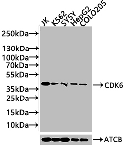
Western Blot
Positive WB detected in: JK whole cell lysate(20μg), K562 whole cell lysate(20μg), SY5Y whole cell lysate(20μg), HepG2 whole cell lysate(20μg), COLO205 whole cell lysate(20μg)
All lanes: CDK6 antibody at 1:1000
Secondary
Goat polyclonal to rabbit IgG at 1/40000 dilution
Predicted band size: 37 kDa
Observed band size: 37 kDa
Exposure time: 120s
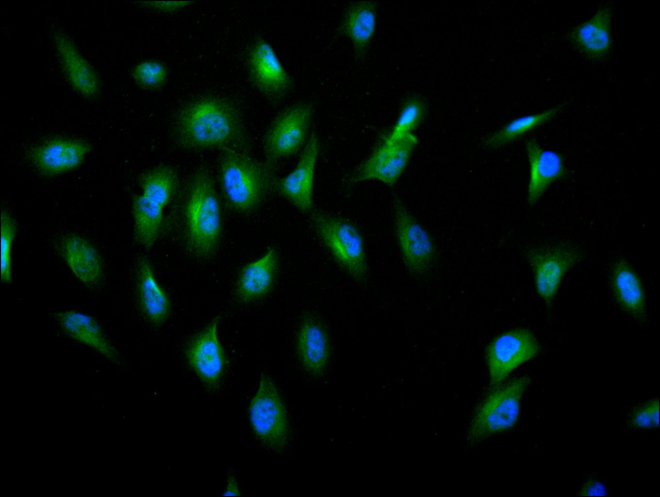
Immunofluorescence staining of U251 cell with CSB-RA555745A0HU at 1:10, counter-stained with DAPI. The cells were fixed in 4% formaldehyde and and permeated by 0.2% TritonX-100 for 15 min. Then 10% normal goat serum to block non-specific protein-protein interactions . The cells were then incubated with the antibody overnight at 4℃. The secondary antibody was Alexa Fluor 488-congugated AffiniPure Goat Anti-Rabbit IgG(H+L).
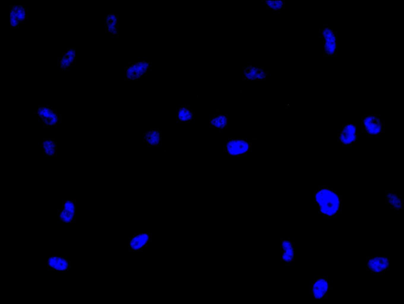
Immunofluorescence staining of U251 cell with 5% goat serum, counter-stained with DAPI. The cells were fixed in 4% formaldehyde and blocked in 10% normal Goat Serum. The cells were then incubated with the antibody overnight at 4C. The secondary antibody was Alexa Fluor 488-congugated AffiniPure Goat Anti-Rabbit IgG(H+L).
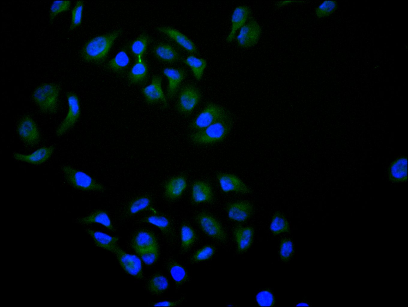
Immunofluorescence staining of Hela cell with CSB-RA555745A0HU at 1:10, counter-stained with DAPI. The cells were fixed in 4% formaldehyde and and permeated by 0.2% TritonX-100 for 15 min. Then 10% normal goat serum to block non-specific protein-protein interactions . The cells were then incubated with the antibody overnight at 4℃. The secondary antibody was Alexa Fluor 488-congugated AffiniPure Goat Anti-Rabbit IgG(H+L).
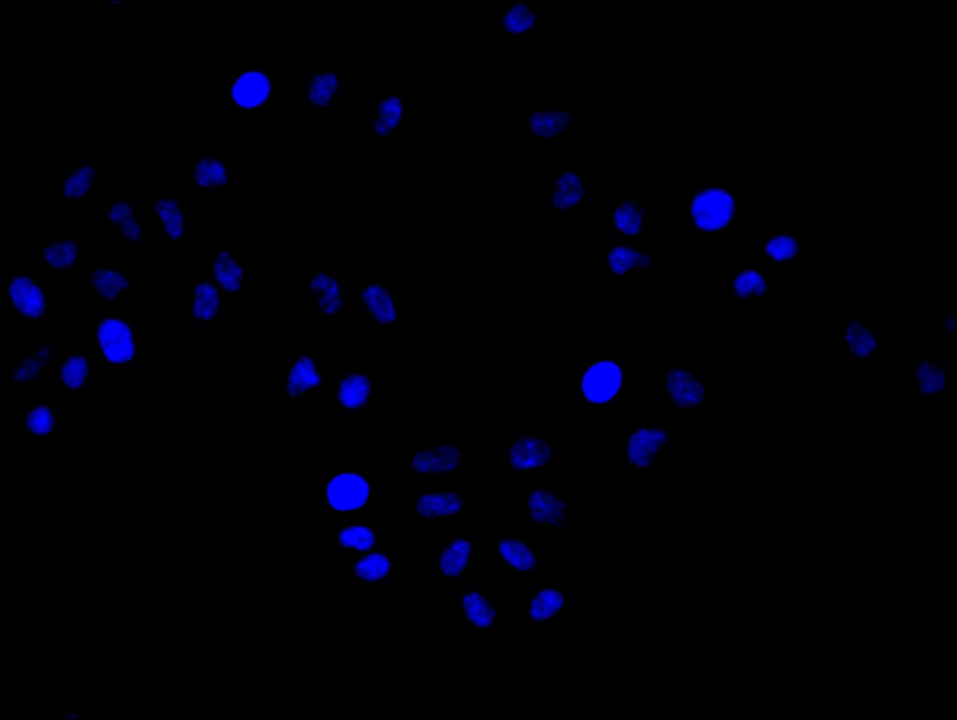
Immunofluorescence staining of Hela cell with 5% goat serum, counter-stained with DAPI. The cells were fixed in 4% formaldehyde and blocked in 10% normal Goat Serum. The cells were then incubated with the antibody overnight at 4C. The secondary antibody was Alexa Fluor 488-congugated AffiniPure Goat Anti-Rabbit IgG(H+L).
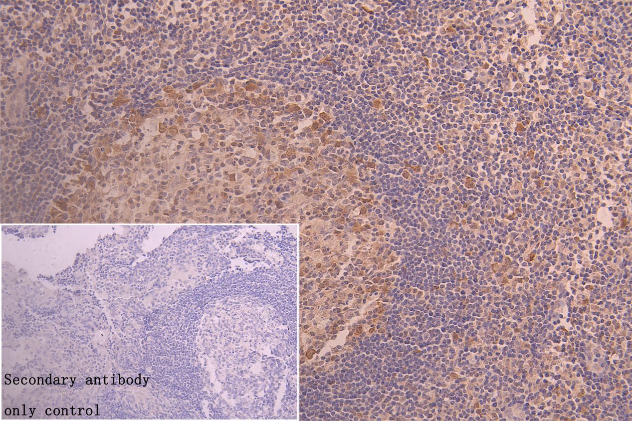
IHC image of CSB-RA555745A0HU diluted at 1:50 and staining in paraffin-embedded human tonsil tissue performed on a Leica BondTM system. After dewaxing and hydration, antigen retrieval was mediated by high pressure in a citrate buffer (pH 6.0). Section was blocked with 10% normal goat serum 30min at RT. Then primary antibody (1% BSA) was incubated at 4°C overnight. The primary is detected by a Goat anti-rabbit polymer IgG labeled by HRP and visualized using 0.05% DAB. Secondary antibody only control: uses 1% BSA instead of primary antibody
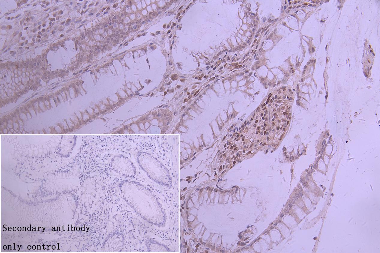
IHC image of CSB-RA555745A0HU diluted at 1:50 and staining in paraffin-embedded human colorectal cancer performed on a Leica BondTM system. After dewaxing and hydration, antigen retrieval was mediated by high pressure in a citrate buffer (pH 6.0). Section was blocked with 10% normal goat serum 30min at RT. Then primary antibody (1% BSA) was incubated at 4°C overnight. The primary is detected by a Goat anti-rabbit polymer IgG labeled by HRP and visualized using 0.05% DAB. Secondary antibody only control: uses 1% BSA instead of primary antibody
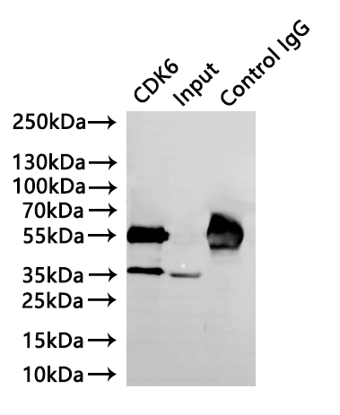
Immunoprecipitating CDK6 in K562whole cell lysate
Lane 1: CSB-RA555745A0HU(3μg)+ K562 whole cell lysate(220μg)
Lane 2: K562 whole cell lysate(30μg)
Lane 3:Rabbit control IgG instead of CSB-RA010605A0HU?in?K562?whole?cell?lysate
For western blotting, a HRP-conjugated Protein G antibody was used as the secondary antibody (1/40000)
CDK6 Antibodies
CDK6 for Homo sapiens (Human)
| 產品貨號 | 產品名稱 | 種屬反應性 | 應用類型 |
|---|---|---|---|
| CSB-PA005074GA01HU | CDK6 Antibody | Human,Mouse,Rat | ELISA,WB |
| CSB-PA549404 | Phospho-CDK6 (Tyr13) Antibody | Human,Mouse | ELISA,WB,IHC,IF |
| CSB-PA267478 | Phospho-CDK6 (Tyr24) Antibody | Human,Mouse | ELISA,WB,IHC,IF |
| CSB-PA090625 | CDK6 (Ab-13) Antibody | Human,Mouse | ELISA,IHC,IF |
| CSB-PA564539 | CDK6 (Ab-24) Antibody | Human,Mouse | ELISA,WB,IHC |
| CSB-PA871419 | CDK6 Antibody | Human,Mouse | ELISA,WB,IHC |
| CSB-PA245607 | CDK6 Antibody | Human | WB, IHC, ELISA |
| CSB-PA005074LA01HU | CDK6 Antibody | Human | ELISA, IHC, IF |
| CSB-PA005074LB01HU | CDK6 Antibody, HRP conjugated | Human | ELISA |
| CSB-PA005074LC01HU | CDK6 Antibody, FITC conjugated | Human | |
| CSB-PA005074LD01HU | CDK6 Antibody, Biotin conjugated | Human | ELISA |
| CSB-RA555745A0HU | CDK6 Recombinant Monoclonal Antibody | Human | ELISA, WB, IHC, IF, IP |
CDK6 Proteins
CDK6 Proteins for Mus musculus (Mouse)
| 產品貨號 | 產品名稱 | 來源 |
|---|---|---|
| CSB-YP005074MO CSB-EP005074MO CSB-BP005074MO CSB-MP005074MO CSB-EP005074MO-B |
Recombinant Mouse Cyclin-dependent kinase 6 (Cdk6) | Yeast E.coli Baculovirus Mammalian cell In Vivo Biotinylation in E.coli |