Nestin 單克隆抗體
日期:2016-12-12 08:56:20
Nestin結構特征
Nestin為第VI類中間絲蛋白,其分子量為 220KD~250KD,位于1號染色體23.1q。Nestin基因含有三個內含子和四個外顯子,三個內含子中有兩個是與神經絲共享的,充分表明Nestin與神經絲來自于一個祖先的可能性。Nestin合成的蛋白分為三個區域,N端(1-7aa)、α螺旋區(8-313)和N端(314-1621aa),α螺旋區被分為四個部分(1A、1B、2A、2B)。
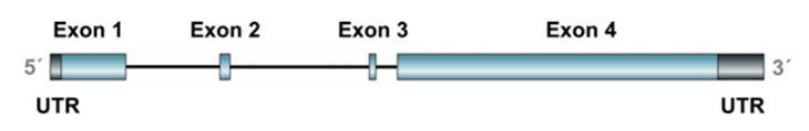
Nestin外顯子與內含子分布結構圖
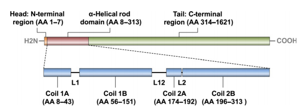
Nestin結構分布圖
Nestin主要功能
(1)參與細胞骨架形成,維持細胞形態;
(2)在有絲分裂過程中促進vimentin的解聚;
(3)維持腦與眼組織的正常發育;
(4)在一些腫瘤中,高表達nestin與腫瘤的浸潤性生長、轉移與不良預后成正相關。
Nestin臨床應用
動物體早期發育過程中,中樞神經系統發育是一個重要事件。Nestin是一種中間絲蛋白,它在哺乳動物神經前體細胞中高表達,已被廣泛用作神經前體細胞的標志分子。因此,nestin的表達情況對分析神經系統的進化具有重要作用,同時也可以作為神經系統病變和損傷的快速敏感診斷指標之一。在成體組織中,nestin只在部分具有分化潛能的細胞中表達,如在皮膚、心血管再生過程中表達增高。此外,nestin在多種腫瘤組織中表達增加,蛋白表達量與腫瘤的惡性程度成正相關,因此nestin在腫瘤組織中的表達水平對于評價腫瘤的生物學行為和患者的預后狀況具有重要意義。
我們生產的nestin單克隆抗體(CSB-MA0157131A0m,100µg/50µg)可以應用于ELISA、WB、IHC、WB、IF、FC檢測。WB可識別U251和SH-Sy5y天然樣本;IHC檢測顯示在扁桃體、腎組織和黑色素瘤中為陽性;在Hela、PC-3、U251的IF檢測中具有陽性信號;在hela和U251細胞的流式細胞檢測中呈現強陽性。
WB:
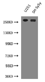
Positive WB detected in:U251 whole cell lysate,SH-Sy5y whole cell lysate
All lanes: NES antibody at 0.6ug/ml
Predicted band size: 260 KDa
Observed band size: 260 KDa
IHC:
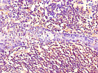
Immunohistochemistry of paraffin-embedded human tonsil tissue using CSB-MA0157131A0m at dilution of 1:100.
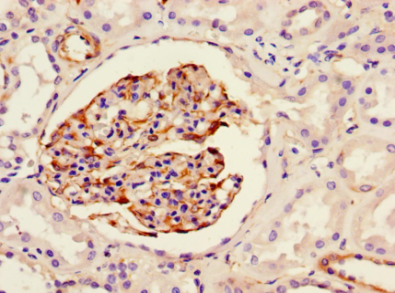
Immunohistochemistry of paraffin-embedded human kidney tissue using CSB-MA0157131A0m at dilution of 1:100.
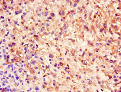
Immunohistochemistry of paraffin-embedded human melanoma using CSB-MA0157131A0m at dilution of 1:100.
IF:
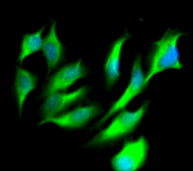
Immunofluorescent analysis of Hela cells using CSB-MA0157131A0m at a dilution of 1:100 and Alexa Fluor 488-congugated AffiniPure Goat Anti-Mouse IgG(H+L)
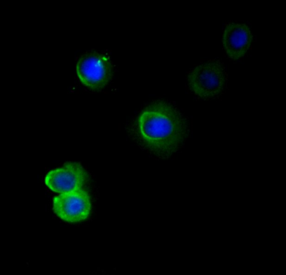
Immunofluorescent analysis of PC-3 cells using CSB-MA0157131A0m at a dilution of 1:100 and Alexa Fluor 488-congugated AffiniPure Goat Anti-Mouse IgG(H+L)
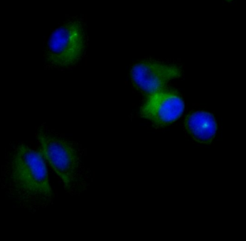
Immunofluorescent analysis of U251 cells using CSB-MA0157131A0m at a dilution of 1:100 and Alexa Fluor 488-congugated AffiniPure Goat Anti-Mouse IgG(H+L)
FC:
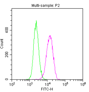
Overlay histogram showing U251 cells stained with CSB-MA0157131A0m (red line). The cells were fixed with 70% ethylalcohol (18h) and then permeabilized with 0.3% Triton X-100 for 2 min. The cells were then incubated in 1x PBS /10% normal goat serum to block non-specific protein-protein interactions followed by the antibody (10µg/1x106cells) for 1 h at 4℃. The secondary antibody used was FITC goat anti-mouse IgG (H+L) at 1/200 dilution for 1 h at 4℃. Isotype control antibody (green line) was mouse IgG1 (10µg/1x106cells) used under the same conditions. Acquisition of >10,000 events was performed.
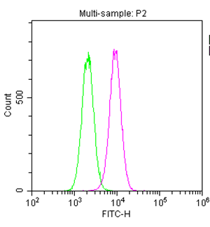
Overlay histogram showing Hela cells stained with CSB-MA0157131A0m (red line). The cells were fixed with 70% ethylalcohol (18h) and then permeabilized with 0.3% Triton X-100 for 2 min. The cells were then incubated in 1x PBS /10% normal goat serum to block non-specific protein-protein interactions followed by the antibody (10µg/1x106cells) for 1 h at 4℃. The secondary antibody used was FITC goat anti-mouse IgG (H+L) at 1/200 dilution for 1 h at 4℃. Isotype control antibody (green line) was mouse IgG1 (10µg/1x106cells) used under the same conditions. Acquisition of >10,000 events was performed.
上一篇: 喂,你竟然是個這樣的TM4SF1
下一篇: 別光顧著羅一笑,白血病你知道多少