TUBB Monoclonal Antibody
-
中文名稱:TUBB 鼠單克隆抗體
-
貨號:CSB-MA025318A0m
-
規(guī)格:¥400
-
圖片:
-
 Western Blot
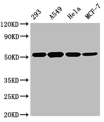
Positive WB detected in: 293 whole cell lysate, A549 whole cell lysate, Hela whole cell lysate, MCF-7 whole cell lysate
All lanes TUBB antibody at 1:5000
Secondary
Goat polyclonal to mouse IgG at 1/50000 dilution
Predicted band size: 55 KDa
Observed band size: 55 KDa
Exposure time:5s -
 Western Blot
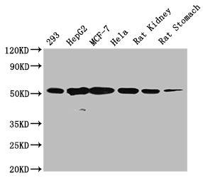
Positive WB detected in: 293 whole cell lysate, HepG2 whole cell lysate, MCF-7 whole cell lysate, Hela whole cell lysate , Rat kidney tissue , Rat stomach tissue
All lanes TUBB antibody at 1:5000
Secondary
Goat polyclonal to mouse IgG at 1/50000 dilution
Predicted band size: 55 KDa
Observed band size: 55 KDa
Exposure time:5min -
 Western Blot
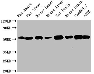
Positive WB detected in: Rat heart tissue ,Rat liver tissue, Mouse heart tissue, Mouse liver tissue, Rat brain tissue, Mouse brain tissue, Raw264.7 whole cell lysate, A375 whole cell lysate
All lanes TUBB antibody at 1:5000
Secondary
Goat polyclonal to mouse IgG at 1/50000 dilution
Predicted band size: 55 KDa
Observed band size: 55 KDa
Exposure time:5min -
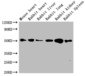 Western Blot
Positive WB detected in: Mouse heart tissue, Rabbit heart tissue, Rabbit liver tissue, Rabbit lung tissue, Rabbit kidney tissue, Rabbit spleen tissue
All lanes TUBB antibody at 1:5000
Secondary
Goat polyclonal to mouse IgG at 1/50000 dilution
Predicted band size: 55 KDa
Observed band size: 55 KDa
Exposure time:5min -
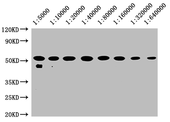 Western Blot
Positive WB detected in: 20μg hela whole cell lysate TUBB antibody at 1:5000, 1:10000, 1:20000, 1:40000, 1:80000, 1:160000, 1:320000, 1:640000
Secondary
Goat polyclonal to mouse IgG at 1/50000 dilution
Predicted band size: 55 KDa
Observed band size: 55 KDa
Exposure time:5min -
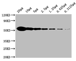 Western Blot
Positive WB detected in: Hela whole cell lysate at 20μg, 10μg, 5μg, 2.5μg, 1.25μg, 0.625μg, 0.3125μg All lanes:TUBB antibody at 1:5000
Secondary
Goat polyclonal to mouse IgG at 1/50000 dilution
Predicted band size: 55 KDa
Observed band size: 55 KDa
Exposure time:5min -
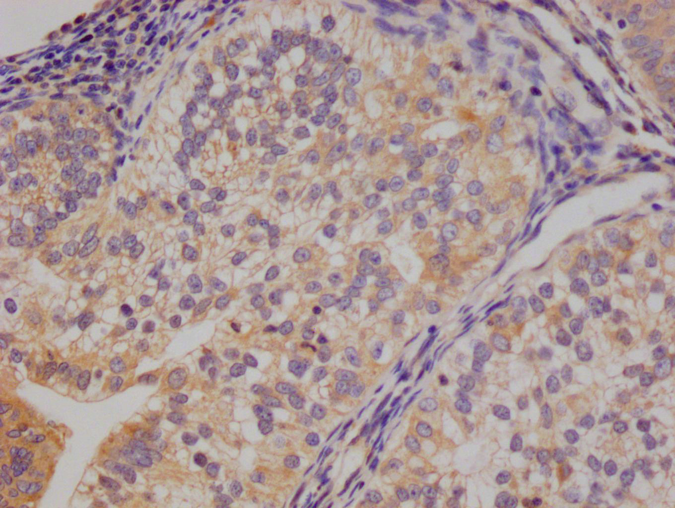
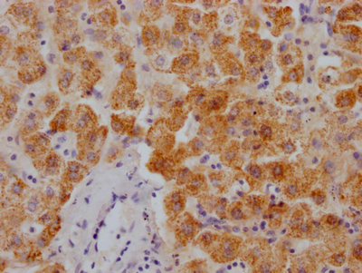
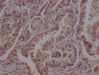 IHC image of CSB-MA025318A0m diluted at 1:200 and staining in paraffin-embedded human lung cancer performed on a Leica BondTM system. After dewaxing and hydration, antigen retrieval was mediated by high pressure in a citrate buffer (pH 6.0). Section was blocked with 10% normal goat serum 30min at 37°C Then primary antibody (1% BSA) was incubated at 4°C overnight. The primary is detected by a Goat anti-Mouse IgG labeled by HRP and visualized using 0.05% DAB.
 -
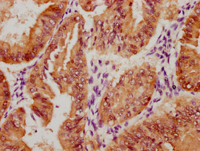
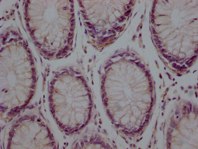 IHC image of CSB-MA025318A0m diluted at 1:200 and staining in paraffin-embedded human colon cancer performed on a Leica BondTM system. After dewaxing and hydration, antigen retrieval was mediated by high pressure in a citrate buffer (pH 6.0). Section was blocked with 10% normal goat serum 30min at 37°C Then primary antibody (1% BSA) was incubated at 4°C overnight. The primary is detected by a Goat anti-Mouse IgG labeled by HRP and visualized using 0.05% DAB.
 -
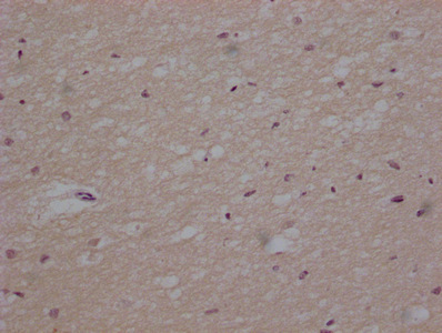 IHC image of CSB-MA025318A0m diluted at 1:200 and staining in paraffin-embedded human brain tissue performed on a Leica BondTM system. After dewaxing and hydration, antigen retrieval was mediated by high pressure in a citrate buffer (pH 6.0). Section was blocked with 10% normal goat serum 30min at 37°C Then primary antibody (1% BSA) was incubated at 4°C overnight. The primary is detected by a Goat anti-Mouse IgG labeled by HRP and visualized using 0.05% DAB.
 -
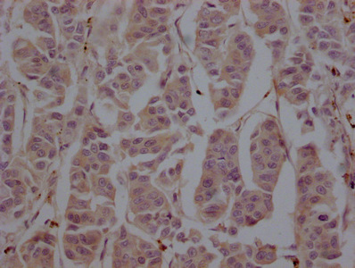 IHC image of CSB-MA025318A0m diluted at 1:200 and staining in paraffin-embedded human breast cancer performed on a Leica BondTM system. After dewaxing and hydration, antigen retrieval was mediated by high pressure in a citrate buffer (pH 6.0). Section was blocked with 10% normal goat serum 30min at 37°C Then primary antibody (1% BSA) was incubated at 4°C overnight. The primary is detected by a Goat anti-Mouse IgG labeled by HRP and visualized using 0.05% DAB.
 -
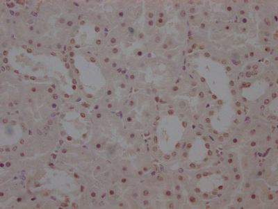
 IHC image of CSB-MA025318A0m diluted at 1:200 and staining in paraffin-embedded human kidney tissue performed on a Leica BondTM system. After dewaxing and hydration, antigen retrieval was mediated by high pressure in a citrate buffer (pH 6.0). Section was blocked with 10% normal goat serum 30min at 37°C Then primary antibody (1% BSA) was incubated at 4°C overnight. The primary is detected by a Goat anti-Mouse IgG labeled by HRP and visualized using 0.05% DAB.
 -
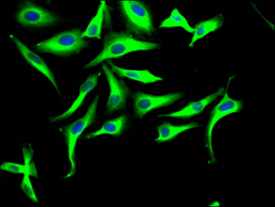
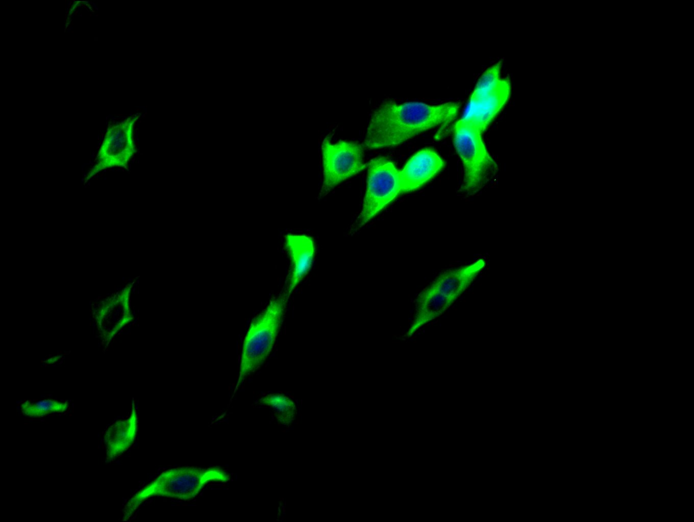 Immunofluorescence staining of NIH/3T3 cells with CSB-MA025318A0m at 1:100, counter-stained with DAPI. The cells were fixed in 4% formaldehyde, permeated by 0.2% TritonX-100, and blocked in 10% normal Goat Serum. The cells were then incubated with the antibody overnight at 4°C. Nuclear DNA was labeled in blue with DAPI. The secondary antibody was FITC-conjugated AffiniPure Goat Anti-Mouse IgG(H+L).
 -
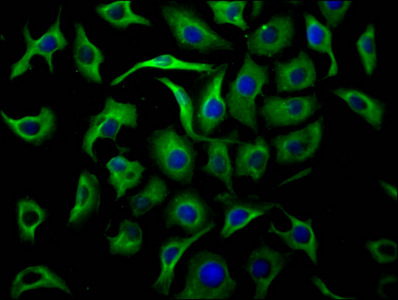 Immunofluorescence staining of A549 cells with CSB-MA025318A0m at 1:100, counter-stained with DAPI. The cells were fixed in 4% formaldehyde, permeated by 0.2% TritonX-100, and blocked in 10% normal Goat Serum. The cells were then incubated with the antibody overnight at 4°C. Nuclear DNA was labeled in blue with DAPI. The secondary antibody was FITC-conjugated AffiniPure Goat Anti-Mouse IgG(H+L).
 -
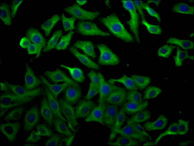
 Immunofluorescence staining of Hela cells with CSB-MA025318A0m at 1:100, counter-stained with DAPI. The cells were fixed in 4% formaldehyde, permeated by 0.2% TritonX-100, and blocked in 10% normal Goat Serum. The cells were then incubated with the antibody overnight at 4°C. Nuclear DNA was labeled in blue with DAPI. The secondary antibody was FITC-conjugated AffiniPure Goat Anti-Mouse IgG(H+L).
 -
 Immunofluorescence staining of HepG2 cells with CSB-MA025318A0m at 1:100, counter-stained with DAPI. The cells were fixed in 4% formaldehyde, permeated by 0.2% TritonX-100, and blocked in 10% normal Goat Serum. The cells were then incubated with the antibody overnight at 4°C. Nuclear DNA was labeled in blue with DAPI. The secondary antibody was FITC-conjugated AffiniPure Goat Anti-Mouse IgG(H+L).
 -
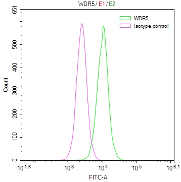
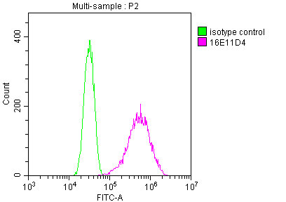
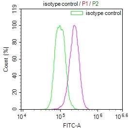
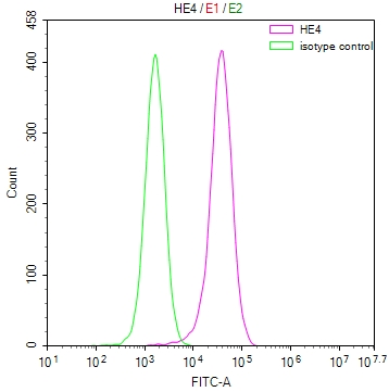
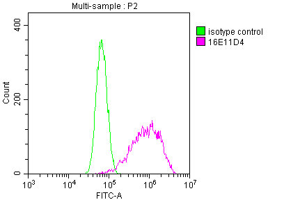 Overlay Peak curve showing A549 cells stained with CSB-MA025318A0m (red line) at 1:200. The cells were fixed in 4% formaldehyde and permeated by 0.2% TritonX-100. Then 10% normal goat serum was Incubated to block non-specific protein-protein interactions followed by the antibody (1μg/1*106cells) for 1 h at 4°C. The secondary antibody used was FITC-conjugated Goat Anti-Mouse IgG(H+L) at 1/100 dilution for 30min at 4°C. Isotype control antibody (green line) was mouse IgG2b (1μg/1*106cells) used under the same conditions. Acquisition of >10,000 events was performed.
 -
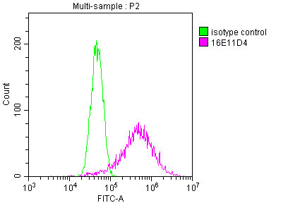
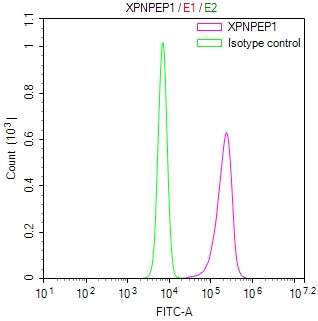
 Overlay Peak curve showing HepG2 cells stained with CSB-MA025318A0m (red line) at 1:200. The cells were fixed in 4% formaldehyde and permeated by 0.2% TritonX-100. Then 10% normal goat serum was Incubated to block non-specific protein-protein interactions followed by the antibody (1μg/1*106cells) for 1 h at 4°C. The secondary antibody used was FITC-conjugated Goat Anti-Mouse IgG(H+L) at 1/100 dilution for 30min at 4°C. Isotype control antibody (green line) was mouse IgG2b (1μg/1*106cells) used under the same conditions. Acquisition of >10,000 events was performed.
 -
 Overlay Peak curve showing MCF-7 cells stained with CSB-MA025318A0m (red line) at 1:200. The cells were fixed in 4% formaldehyde and permeated by 0.2% TritonX-100. Then 10% normal goat serum was Incubated to block non-specific protein-protein interactions followed by the antibody (1μg/1*106cells) for 1 h at 4°C. The secondary antibody used was FITC-conjugated Goat Anti-Mouse IgG(H+L) at 1/100 dilution for 30min at 4°C. Isotype control antibody (green line) was mouse IgG2b (1μg/1*106cells) used under the same conditions. Acquisition of >10,000 events was performed.
 -
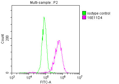 Overlay Peak curve showing NIH/3T3 cells stained with CSB-MA025318A0m (red line) at 1:200. The cells were fixed in 4% formaldehyde and permeated by 0.2% TritonX-100. Then 10% normal goat serum was Incubated to block non-specific protein-protein interactions followed by the antibody (1μg/1*106cells) for 1 h at 4°C. The secondary antibody used was FITC-conjugated Goat Anti-Mouse IgG(H+L) at 1/100 dilution for 30min at 4°C. Isotype control antibody (green line) was mouse IgG2b (1μg/1*106cells) used under the same conditions. Acquisition of >10,000 events was performed.
 -
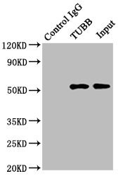 Immunoprecipitating TUBB in Hela whole cell lysate
Lane 1: Mouse control IgG instead of CSB-MA025318A0m in Hela whole cell lysate.
Lane 2: CSB-MA025318A0m (2μg) + Hela whole cell lysate (500μg)
Lane 3: Hela whole cell lysate (5μg)
For western blotting, the blot was detected with CSB-MA025318A0m at 1:2000, and a HRP-conjugated Protein G antibody was used as the secondary antibody at 1:5000
-
-
其他:
產(chǎn)品詳情
-
產(chǎn)品描述:TUBB單克隆抗體(CUSABIO貨號:CSB-MA025318A0m)是一款針對β-微管蛋白(TUBB)的高特異性科研試劑,該靶標(biāo)作為細(xì)胞骨架的關(guān)鍵組分,在細(xì)胞分裂、胞內(nèi)運輸及形態(tài)維持中發(fā)揮重要作用。本產(chǎn)品采用雜交瘤技術(shù)制備,經(jīng)嚴(yán)格質(zhì)控驗證其與人、大鼠、兔、小鼠等多物種TUBB蛋白的高親和力結(jié)合特性,可兼容ELISA、Western Blot、免疫組化(IHC)、免疫熒光(IF)、流式細(xì)胞術(shù)(FC)及免疫沉淀(IP)六大實驗平臺。在神經(jīng)生物學(xué)領(lǐng)域,該抗體適用于神經(jīng)元微管網(wǎng)絡(luò)的可視化分析;在腫瘤研究中可用于檢測癌細(xì)胞分裂過程中微管動態(tài)變化;在發(fā)育生物學(xué)中可追蹤胚胎發(fā)育階段的細(xì)胞骨架重構(gòu)。其寬泛的物種兼容性特別適合跨物種比較研究,而多重應(yīng)用場景的覆蓋為細(xì)胞周期調(diào)控、藥物靶點篩選及細(xì)胞器運輸機制等課題提供可靠工具。作為β-微管蛋白單克隆抗體中的明星產(chǎn)品,CUSABIO貨號:CSB-MA025318A0m憑借卓越的批間穩(wěn)定性和高信噪比特性,已成為細(xì)胞骨架研究、微管相關(guān)蛋白互作分析及分子病理學(xué)機制探索的首選試劑。
-
Uniprot No.:
-
別名:Beta 4 tubulin antibody; Beta 5 tubulin antibody; beta Ib tubulin antibody; Beta1 tubulin antibody; Class I beta tubulin antibody; M40 antibody; MGC117247 antibody; MGC16435 antibody; OK/SW cl.56 antibody; OK/SWcl.56 antibody; TBB5_HUMAN antibody; TUBB 1 antibody; TUBB 2 antibody; TUBB 5 antibody; TUBB antibody; TUBB1 antibody; TUBB2 antibody; TUBB5 antibody; tubulin beta 1 chain antibody; Tubulin beta 2 chain antibody; tubulin beta 5 chain antibody; Tubulin beta chain antibody; Tubulin beta class I antibody; tubulin beta polypeptide antibody; Tubulin beta-5 chain antibody
-
宿主:Mouse
-
反應(yīng)種屬:Human, Rat, Rabbit, Mouse
-
免疫原:GAGNNWAKGHYTEGA synthetic peptide conjugate to KLH
-
標(biāo)記方式:Non-conjugated
-
克隆類型:Monoclonal Antibody
-
抗體亞型:IgG2b
-
純化方式:>95%, Protein A purified
-
克隆號:16E11D4
-
濃度:It differs from different batches. Please contact us to confirm it.
-
保存緩沖液:Preservative: 0.03% Proclin 300
Constituents: 50% Glycerol, 0.01M PBS, PH 7.4 -
產(chǎn)品提供形式:Liquid
-
應(yīng)用范圍:ELISA, WB, IHC, IF, FC, IP
-
推薦稀釋比:
Application Recommended Dilution WB 1:5000-1:640000 IHC 1:100-1:300 IF 1:50-1:200 FC 1:100-1:300 IP 1μl-2μl -
Protocols:
-
儲存條件:Upon receipt, store at -20°C or -80°C. Avoid repeated freeze.
-
貨期:Basically, we can dispatch the products out in 1-3 working days after receiving your orders. Delivery time maybe differs from different purchasing way or location, please kindly consult your local distributors for specific delivery time.
-
用途:For Research Use Only. Not for use in diagnostic or therapeutic procedures.
產(chǎn)品評價

Most popular with customers
-
-
YWHAB Recombinant Monoclonal Antibody
Applications: ELISA, WB, IHC, IF, FC
Species Reactivity: Human, Mouse, Rat
-
Phospho-YAP1 (S127) Recombinant Monoclonal Antibody
Applications: ELISA, WB, IHC
Species Reactivity: Human
-
-
-
-
-