UBA2 Recombinant Monoclonal Antibody
-
中文名稱:UBA2 Recombinant Monoclonal Antibody
-
貨號:CSB-RA266118A0HU
-
規格:¥1320
-
圖片:
-
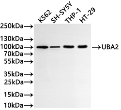 Western Blot
Positive WB detected in: K562 whole cell lysate(30μg), SH-SY5Y whole cell lysate(30μg), THP-1 whole cell lysate(30μg), HT-29 whole cell lysate(30μg)
All lanes: UBA2 antibody at 1:1000
Secondary
Goat polyclonal to rabbit IgG at 1/40000 dilution
Predicted band size: 71 kDa
Observed band size: 100 kDa
Exposure time:2min -
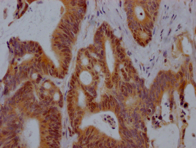
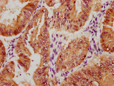
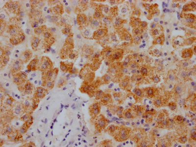
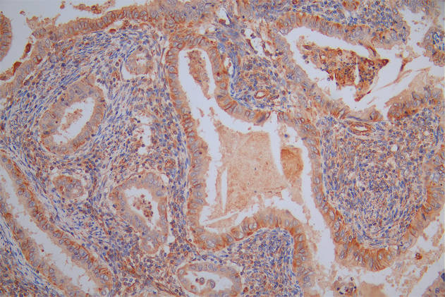
 IHC image of CSB-RA266118A0HU diluted at 1:100 and staining in paraffin-embedded human glioma cancer performed on a Leica BondTM system. After dewaxing and hydration, antigen retrieval was mediated by high pressure in a citrate buffer (pH 6.0). Section was blocked with 10% normal goat serum 30min at RT. Then primary antibody (1% BSA) was incubated at 4°C overnight. The primary is detected by a Goat anti-rabbit polymer IgG labeled by HRP and visualized using 0.05% DAB.
 -
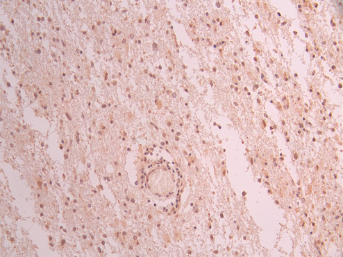
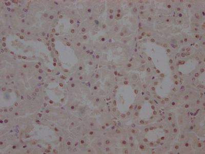
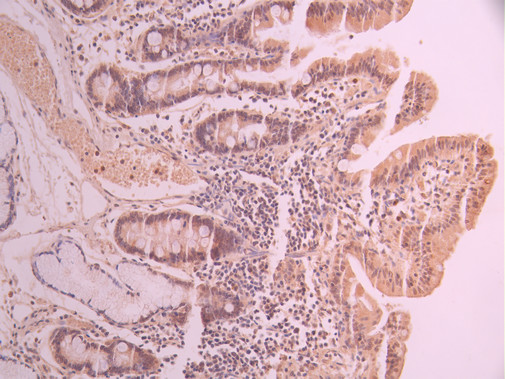 IHC image of CSB-RA266118A0HU diluted at 1:100 and staining in paraffin-embedded human small intestine tissue performed on a Leica BondTM system. After dewaxing and hydration, antigen retrieval was mediated by high pressure in a citrate buffer (pH 6.0). Section was blocked with 10% normal goat serum 30min at RT. Then primary antibody (1% BSA) was incubated at 4°C overnight. The primary is detected by a Goat anti-rabbit polymer IgG labeled by HRP and visualized using 0.05% DAB.
 -
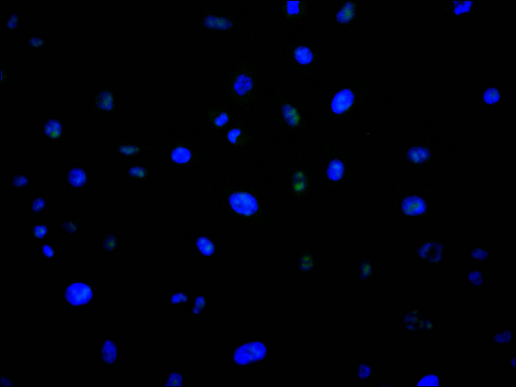 Immunofluorescence staining of A549 cell with CSB-RA266118A0HU at 1:50, counter-stained with DAPI. The cells were fixed in 4% formaldehyde, permeabilized using 0.2% Triton X-100 and blocked in 10% normal Goat Serum. The cells were then incubated with the antibody overnight at 4°C. The secondary antibody was Alexa Fluor 488-congugated AffiniPure Goat Anti-Rabbit IgG(H+L).
 -
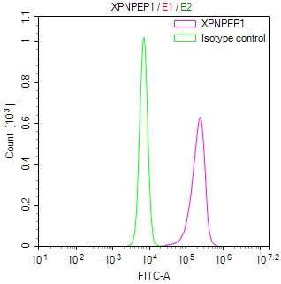
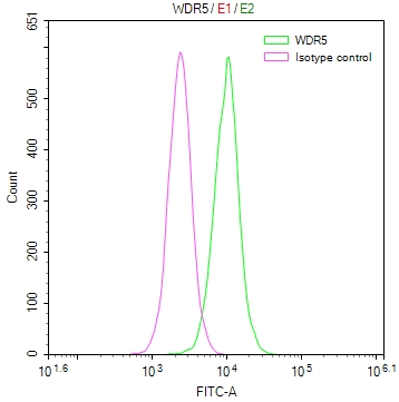
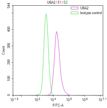 Overlay Peak curve showing jurkat cells stained with CSB-RA266118A0HU (red line) at 1:100. The cells were fixed in 4% formaldehyde and permeated by 0.2% TritonX-100 for?10min. Then 10% normal goat serum to block non-specific protein-protein interactions followed by the antibody (1ug/1*106cells) for 45min at 4℃. The secondary antibody used was FITC-conjugated goat anti-rabbit IgG (H+L) at 1/200 dilution for 35min at 4℃.Control antibody (green line) was Rabbit IgG (1ug/1*106cells) used under the same conditions. Acquisition of >10,000 events was performed.
-
-
其他:
產品詳情
-
Uniprot No.:
-
基因名:
-
別名:SUMO-activating enzyme subunit 2 (EC 2.3.2.-) (Anthracycline-associated resistance ARX) (Ubiquitin-like 1-activating enzyme E1B) (Ubiquitin-like modifier-activating enzyme 2), UBA2, SAE2 UBLE1B
-
反應種屬:Human
-
免疫原:A synthesized peptide from human UBA2 protein
-
免疫原種屬:Homo sapiens (Human)
-
標記方式:Non-conjugated
-
克隆類型:Monoclonal
-
抗體亞型:Rabbit IgG
-
純化方式:Affinity-chromatography
-
克隆號:6E11
-
濃度:It differs from different batches. Please contact us to confirm it.
-
保存緩沖液:Preservative: 0.03% Proclin 300
Constituents: 50% Glycerol, 0.01M PBS, PH 7.4 -
產品提供形式:Liquid
-
應用范圍:ELISA, WB, IHC, IF, FC
-
推薦稀釋比:
Application Recommended Dilution WB 1:500-1:2000 IHC 1:50-1:200 IF 1:50-1:200 FC 1:50-1:200 -
Protocols:
-
儲存條件:Upon receipt, store at -20°C or -80°C. Avoid repeated freeze.
-
貨期:Basically, we can dispatch the products out in 1-3 working days after receiving your orders. Delivery time maybe differs from different purchasing way or location, please kindly consult your local distributors for specific delivery time.
-
用途:For Research Use Only. Not for use in diagnostic or therapeutic procedures.
產品評價

相關產品
靶點詳情
-
功能:The heterodimer acts as an E1 ligase for SUMO1, SUMO2, SUMO3, and probably SUMO4. It mediates ATP-dependent activation of SUMO proteins followed by formation of a thioester bond between a SUMO protein and a conserved active site cysteine residue on UBA2/SAE2.
-
基因功能參考文獻:
- Calcium/calpain-induced cleavage of the SAE2 leads to sumoylation inhibition reslting in bacillary dysentery. PMID: 29231810
- We propose that disturbance of the SUMOylation pathway, mediated by pathogenic variants in UBA2, is a novel mechanism for aplasia cutis congenita and other phenotypic abnormalities. PMID: 28110515
- Here, we show that hHR23A utilizes both the UBA2 and XPCB domains to form a stable complex with Vpr, linking Vpr directly to cellular DNA repair pathways and their probable exploitation by the virus. PMID: 24318982
- This study has identified the mechanism used to localize SAE to the nucleus. PMID: 23095757
- Data indicate the role of anti-SUMO activating enzyme SAE1 and SAE2 antibody as marker of dermatomyositis. PMID: 22884621
- Data show that the SAE2 subunit of the small ubiquitin-like modifier (SUMO) E1 is autoSUMOylated at residue Lys-236, and SUMOylation was catalyzed by Ubc9 at several additional Lys residues surrounding the catalytic Cys-173 of SAE2. PMID: 22403398
- loss of SAE1/2 activity drives synthetic lethality with Myc; inactivation of SAE2 leads to mitotic catastrophe and cell death upon Myc hyperactivation; findings in Myc-high breast cancers suggest low tumor SAE1 and SAE2 correlates metastasis-free survival PMID: 22157079
- The mammalian E1 subunits can be imported separately, identify nuclear localization signals (NLSs) in Aos1 and in Uba2, and demonstrate that their import is mediated by importin alpha/beta in vitro and in intact cells. PMID: 21209321
- structures of heterodimeric Sae1/Sae2-Mg.ATP and Sae1/Sae2-SUMO-1-Mg.ATP complexes PMID: 15660128
- UBA2 stabilizes APOBEC3G by preventing ubiquitin chain elongation and proteasome-mediated proteolysis. PMID: 18680593
顯示更多
收起更多
-
亞細胞定位:Cytoplasm. Nucleus. Note=Shuttles between the cytoplasm and the nucleus, sumoylation is required either for nuclear translocation or nuclear retention.
-
蛋白家族:Ubiquitin-activating E1 family
-
數據庫鏈接:
Most popular with customers
-
Phospho-YAP1 (S127) Recombinant Monoclonal Antibody
Applications: ELISA, WB, IHC
Species Reactivity: Human
-
-
-
-
-
-
-