LAG3 Monoclonal Antibody
-
中文名稱:LAG3鼠單克隆抗體
-
貨號:CSB-MA012719A0m
-
規(guī)格:¥1320
-
圖片:
-
 Western Blot
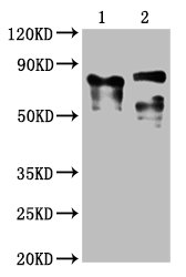
Positive WB detected in: 1 lane: Recombinant proteins with LAG3; 2 lane: 293F whole cell lysate transfected with LAG3
All lanes: LAG3 antibody at 1:2000
Secondary
Goat polyclonal to mouse IgG at 1/50000 dilution
Predicted band size: 76, 88 KDa
Observed band size: 76, 88 KDa
Exposure time:5min -
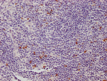
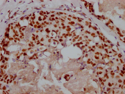
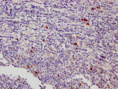 IHC image of CSB-MA012719A0m diluted at 1:500 and staining in paraffin-embedded human tonsil tissue performed on a Leica BondTM system. After dewaxing and hydration, antigen retrieval was mediated by high pressure in a citrate buffer (pH 6.0). Section was blocked with 10% normal goat serum 30min at 37°C Then primary antibody (1% BSA) was incubated at 4°C overnight. The primary is detected by a Goat anti-Mouse IgG labeled by HRP and visualized using 0.05% DAB.
 -
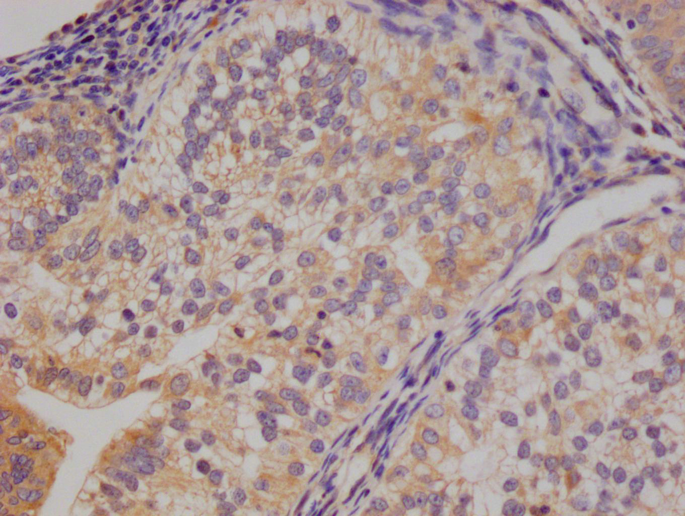
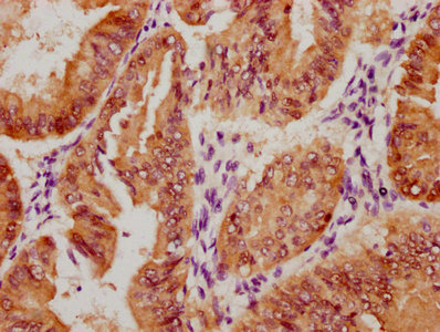
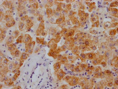
 IHC image of CSB-MA012719A0m diluted at 1:500 and staining in paraffin-embedded human appendix tissue performed on a Leica BondTM system. After dewaxing and hydration, antigen retrieval was mediated by high pressure in a citrate buffer (pH 6.0). Section was blocked with 10% normal goat serum 30min at 37°C Then primary antibody (1% BSA) was incubated at 4°C overnight. The primary is detected by a Goat anti-Mouse IgG labeled by HRP and visualized using 0.05% DAB.
 -
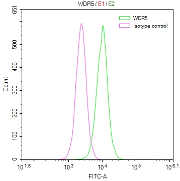
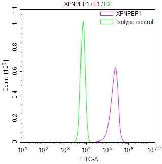
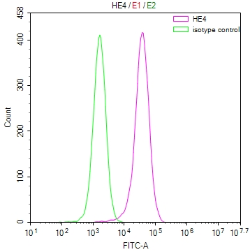
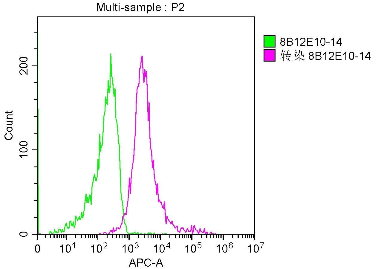 Overlay Peak curve showing A549 cells stained with CSB-MA012719A0m (red line) at 1:100. The cells were incubated in 10% normal goat serum to block non-specific protein-protein interactions followed by the antibody (1μg/1*106cells) for 1h at 4°C. The secondary antibody used was FITC-conjugated Goat Anti-Mouse IgG(H+L) at 1/100 dilution for 30min at 4°C. Isotype control antibody (green line) was mouse IgG1 (1μg/1*106cells) used under the same conditions. Acquisition of >10,000 events was performed.
-
-
其他:
產(chǎn)品詳情
-
產(chǎn)品描述:CUSABIO貨號:CSB-MA012719A0m LAG3單克隆抗體是針對免疫檢查點蛋白LAG3研發(fā)的高特異性科研試劑,適用于ELISA、Western Blot、免疫組化和流式細胞術(shù)等多種實驗平臺。LAG3(淋巴細胞激活基因3)作為重要的免疫調(diào)節(jié)分子,主要表達于活化的T細胞表面,通過與MHC II類分子相互作用抑制T細胞活化,在腫瘤免疫逃逸和自身免疫性疾病中發(fā)揮關(guān)鍵作用,現(xiàn)已成為癌癥免疫治療領(lǐng)域的熱門研究靶點。本產(chǎn)品采用雜交瘤技術(shù)制備,具有優(yōu)異的人源靶標結(jié)合特異性,經(jīng)多批次質(zhì)控驗證其與重組及天然LAG3蛋白的高親和力,適用于檢測細胞裂解液、組織樣本及外周血單核細胞中的LAG3表達水平。科研人員可將其用于腫瘤微環(huán)境分析、免疫檢查點抑制劑作用機制研究、免疫細胞功能評估等前沿領(lǐng)域,為探索LAG3在腫瘤免疫治療中的調(diào)控網(wǎng)絡及開發(fā)新型聯(lián)合治療方案提供可靠工具。該抗體特別適合開展實體瘤免疫組化分析、T細胞活化狀態(tài)流式檢測以及藥物篩選相關(guān)的蛋白印跡實驗,是免疫腫瘤學研究和轉(zhuǎn)化醫(yī)學研究的理想選擇。
-
產(chǎn)品名稱:Mouse anti-Homo sapiens (Human) LAG3 Monoclonal antibody
-
Uniprot No.:
-
基因名:
-
別名:CD223 antibody; CD223 antigen antibody; FDC protein antibody; LAG-3 antibody; Lag3 antibody; LAG3_HUMAN antibody; Lymphocyte activating 3 antibody; Lymphocyte activation gene 3 antibody; Lymphocyte activation gene 3 protein antibody; Protein FDC antibody
-
宿主:Mouse
-
反應種屬:Human
-
免疫原:Recombinant Human Lymphocyte activation gene 3 protein (29-450AA)
-
免疫原種屬:Homo sapiens (Human)
-
標記方式:Non-conjugated
-
克隆類型:Monoclonal
-
抗體亞型:IgG1
-
純化方式:>95%, Protein G purified
-
克隆號:8B12E10
-
濃度:It differs from different batches. Please contact us to confirm it.
-
保存緩沖液:Preservative: 0.03% Proclin 300
Constituents: 50% Glycerol, 0.01M PBS, PH 7.4 -
產(chǎn)品提供形式:Liquid
-
應用范圍:ELISA, WB, IHC, FC
-
推薦稀釋比:
Application Recommended Dilution WB 1:1000-1:5000 IHC 1:200-1:1000 FC 1:50-1:200 -
Protocols:
-
儲存條件:Upon receipt, store at -20°C or -80°C. Avoid repeated freeze.
-
貨期:Basically, we can dispatch the products out in 1-3 working days after receiving your orders. Delivery time maybe differs from different purchasing way or location, please kindly consult your local distributors for specific delivery time.
-
用途:For Research Use Only. Not for use in diagnostic or therapeutic procedures.
產(chǎn)品評價

相關(guān)產(chǎn)品
靶點詳情
-
功能:Lymphocyte activation gene 3 protein: Inhibitory receptor on antigen activated T-cells. Delivers inhibitory signals upon binding to ligands, such as FGL1. FGL1 constitutes a major ligand of LAG3 and is responsible for LAG3 T-cell inhibitory function. Following TCR engagement, LAG3 associates with CD3-TCR in the immunological synapse and directly inhibits T-cell activation. May inhibit antigen-specific T-cell activation in synergy with PDCD1/PD-1, possibly by acting as a coreceptor for PDCD1/PD-1. Negatively regulates the proliferation, activation, effector function and homeostasis of both CD8(+) and CD4(+) T-cells. Also mediates immune tolerance: constitutively expressed on a subset of regulatory T-cells (Tregs) and contributes to their suppressive function. Also acts as a negative regulator of plasmacytoid dendritic cell (pDCs) activation. Binds MHC class II (MHC-II); the precise role of MHC-II-binding is however unclear.; May function as a ligand for MHC class II (MHC-II) on antigen-presenting cells (APC), promoting APC activation/maturation and driving Th1 immune response.
-
基因功能參考文獻:
- Results demonstrated that the expression of LAG3 in colorectal carcinoma tissue was significantly increased compared with the paracancerous tissue. It was mainly expressed at the edge of tumor tissues suggesting that LAG3 was expressed on tumorinfiltrating lymph node cells but not in colorectal cancer cells. PMID: 30272332
- These results together suggested that LAG-3 is a marker of CD4(+) T cells with regulatory function; at the same time, LAG-3 might have limited the full suppressive potential of Treg cells. PMID: 29671649
- Among the 89 patients, CD274, LAG3, and IDO1 expressions in TIICs were observed in 68.6% (61 cases), 13.5% (12), and 28.1% (25) of patients, respectively. CD274, CTLA4, and IDO1 were expressed in tumor cells of 24.7% (22 cases), 4.5% (4), and 72.0% (64) of patients, respectively. PMID: 29520442
- LAG-3+ iTILs are enriched in estrogen receptor-negative breast cancers and represent an independent favorable prognostic factor. In addition, a high proportion of PD-1/PD-L1+ tumors are co-infiltrated with LAG-3+ TILs. PMID: 29045526
- Squamous cell carcinomas escape immune surveillance via inducing chronic activation and exhaustion of CD8+ T Cells co-expressing PD-1 and LAG-3 inhibitory receptors. PMID: 27835902
- Data suggest that blocking LAG3-MHC class II interactions is a potential therapeutic target in chronic lymphocytic leukemia. PMID: 28154084
- LAG-3 expression was correlated with expression of PD-1 on TILs and expression of PD-L1 on tumor cells. Higher expression of LAG-3 on TILs was significantly correlated with higher expression of PD-1 on TILs and higher expression of PD-L1 on tumor cells. PMID: 28132868
- the upregulation of others syncytial molecules, including LAG3, CTLA4, CD28 and CD3, assisting the formation of syncytia with APC cells. PMID: 27108398
- LAG3-expressing CD4(+)CD25(-) T cells represent another regulatory immune cell type with potential to interfere with anti-tumor immunity. PMID: 28935468
- Overexpression of lymphocyte activation gene-3 inhibits regulatory T cell responses in osteoarthritis PMID: 28800255
- Egr2-driven cell surface proteins LAG-3 and 4-1BB can identify dysfunctional tumor antigen-specific CD8(+) TIL. PMID: 28115575
- our data indicate that the evaluation of stromal TILs remains the most reliable immune prognostic marker in TNBC, and support the clinical evaluation of anti-PD-1/PD-L1 and anti-LAG-3 in a subset of patients with TNBC who have concurrent expression of both checkpoint receptors. PMID: 27912781
- this review provides the translational rationale for targeting LAG3, as well as historical and current state of clinical trials utilizing LAG3 cancer immunomodulators PMID: 28258692
- this study shows that LAG3 expressed on myeloid leukaemia cells possess the capacity to facilitate functional exhaustion in T helper cells PMID: 27565576
- epigenetic modification on LAG-3 increased LAG-3(+) T cells and its immune regulatory roles in chronic osteomyelitis progression PMID: 28028751
- Immune checkpoint proteins are co-inhibitory factors that can diminish the antigen-specific immune responses by attenuating the regulatory role of cytotoxic T-lymphocyte-associated protein 4, programmed cell death-1, lymphocyte-activation gene 3, and T-cell immunoglobulin mucin-3. PMID: 28349816
- LAG3 binds alpha-synuclein preformed fibrils (PFF) with high affinity (dissociation constant = 77 nanomolar), whereas the alpha-syn monomer exhibited minimal binding. alpha-Syn-biotin PFF binding to LAG3 initiated alpha-syn PFF endocytosis, transmission, and toxicity. PMID: 27708076
- An IL-27/Lag3 axis enhances Foxp3+ regulatory T cell-suppressive function and therapeutic efficacy. PMID: 26013006
- LAG-3 is highly expressed in peripheral blood CD8+ T cells in chronic HBV-infected patients. PMID: 27053622
- iNKT cytokine production is profoundly altered by both HIV infection and treatment, and LAG-3, but not PD-1, expression is associated with a reduction in iNKT IFNgamma production. PMID: 25810006
- NFKB1, CD27, LAG3 and IKZF3 are new susceptibility genes for psoriasis. PMID: 25006012
- The elevated expression of LAG-3 at the genital tract suggests it may regulate T-cell activation, and identify cells susceptible to HIV infection. The enrichment of LAG-3 on double negative T cells suggests LAG-3 may contribute to the immunoregulatory activity of these cells. PMID: 25154740
- the LAG-3/MHC class II pathway may synergize with PD-1/PD ligand to enhance T cell-mediated immune responses. PMID: 25780040
- LAG-3 trafficking from lysosomal compartments to the cell surface is dependent on the cytoplasmic domain through protein kinase C signaling in activated T cells. PMID: 25108024
- These results suggest that LAG-3-mediated activation of plasmacytoid dendritic cells takes place in vivo at tumor sites, and it is in part responsible for directing an immune-suppressive environment. PMID: 24441096
- Expression of LAG-3 is coincident with the impaired effector function of HBV-specific CD8(+) T cell in HCC patients. PMID: 23261718
- our data suggest that the LAG-3-MHC II interaction could be viewed as a bidirectional immune escape pathway in melanoma PMID: 21441454
- Multiple myeloma was associated with 2 SNPs in intron regions of LAG3 within 20 k base pairs 5' upstream of the candidate CD4 gene. The variant in LAG3 gene itself may play a role in susceptibility of MM. PMID: 20568250
- Analysis identified a gene-set (LAG3, LPL, ZAP70) whose overexpression is assigned to unmutated immunoglobulin variable heavy chain region with 90% specificity. PMID: 20228263
- LAG-3 defines an active CD4(+)CD25(high)Foxp3(+) regulatory T cell subset whose frequency is enhanced in the PBMCs of patients with cancer PMID: 20421648
- Tumor-infiltrating NY-ESO-1-specific CD8+ T cells are negatively regulated by LAG-3 and PD-1 in human ovarian cancer. PMID: 20385810
- MHC class II-mediated signals induced by the natural ligand, LAG-3, lead to complete maturation of monocyte-derived dendritic cells, which acquire the capacity to trigger naive T cells and drive polarized Th1 responses. PMID: 11937541
- LAG-3 induces rapid protein phosphorylation of phospholipase Cgamma2 and p72syk as well as activation of phosphatidyl inositol 3-kinase/Akt, p42/44 extracellular signal-regulated protein kinase, and p38 mitogen-activated protein kinase pathways. PMID: 12775570
- LAG3 may mediate sphingolipid metabolism PMID: 12825348
- LAG-3 activates antigen-presenting cells through MHC class II signalling, leading to increased antigen-specific T-cell responses in vivo. PMID: 14644131
- regulatory T cells and LAG-3 have pivotal roles in the suppression of EBV-specific cell-mediated immunity in Hodgkin lymphoma PMID: 16757686
- The result, including a total of 2640 MS patients and 2194 controls shows no significant association with CD4 and LAG3 and MS. We conclude that these genes are of minor importance in regard of genetic predisposition to the MS. PMID: 17020785
- Soluble human recombinant fusion protein (hLAG-3Ig) used in vitro as a single maturation agent induces phenotypic maturation of monocyte-derived dendritic cells and promotes the production of chemokines and TNF-alpha inflammatory cytokine. PMID: 18322184
顯示更多
收起更多
-
亞細胞定位:[Lymphocyte activation gene 3 protein]: Cell membrane; Single-pass type I membrane protein.; [Secreted lymphocyte activation gene 3 protein]: Secreted.
-
組織特異性:Primarily expressed in activated T-cells and a subset of natural killer (NK) cells.
-
數(shù)據(jù)庫鏈接:
Most popular with customers
-
-
Phospho-YAP1 (S127) Recombinant Monoclonal Antibody
Applications: ELISA, WB, IHC
Species Reactivity: Human
-
-
-
-
-
-